Int J Pharm Pharm Sci, Vol 6, Issue 9, 86-90Original Article
EVALUATION OF CARDIO PROTECTIVE ACTIVITY OF GALANGIN AGAINST DOXORUBICIN INDUCED CARDIOMYOPATHY
RAVICHANDRA V, HANUMANTHARAYAPPA B*, MADHAVA REDDY PAPASANI V
Gautham College of Pharmacy, No.32, 3rd "A" Cross, Kanakanagar, RT Nagar Post, Bangalore 560032, Karnataka, India.
Email: findraya@gmail.com
Received: 17 May 2014 Revised and Accepted: 10 Aug 2014
ABSTRACT
Objective: The present study was designed to investigate the cardioprotective potential of Galangin on Doxorubicin induced cardiotoxicity in rats.
Methods: Albino rats used in this experiment were pretreated with vehicle, Galangin (100 & 200µg/kg) and Vit-C (20 mg/kg) for 28 days. On 25th day, a single dose of Doxorubicin (10 mg/kg, i. p) was administered to groups. After 72 h of Doxorubicin administration, ECG, serum and tissues biomarkers were evaluated. Histopathological examination of the heart was performed.
Results: Doxorubicin treated rats exhibited abnormal ECG pattern followed by significant increase in CK-MB, LDH, SGOT, SGPT and LPO level and decrease in GSH, CAT, TT when compared to control rats. Pretreatment with different doses of Galangin and Vit-C significantly reduced the serum biomarkers and increased the tissue antioxidant level when compared to Doxorubicin alone treated groups. Moreover, pretreatment also improved Doxorubicin induced changes in ECG pattern and histopathology of heart.
Conclusion: We conclude that the present study provides experimental evidence that Galangin has strong antioxidant activity, and it can maintain cell membrane integrity, and ameliorate oxidative stress induced by high-dose of Doxorubicin administration. These findings might be helpful to understand the beneficial effects of Galangin against myocardial injury although further study is needed to confirm its mechanism.
Keywords: Doxorubicin, Cardiotoxicity, Galangin, Vitamin C, Cardioprotective.
INTRODUCTION
Among the many health predictions for the new millennium, the most alarming is that of cardiovascular disease (CVD), heart diseases and stroke topping the list for death and disability [1]. The major risk factors associated with CHD and stroke are tobacco and alcohol abuse, high blood pressure (hypertension), high cholesterol, obesity, physical inactivity, and unhealthy diets contributing to high prevalence across the world [2]. Current projections suggest that by the year 2020 India will have the largest CVD burden in the world. One fifth of the deaths in India are from CHD. By the year 2020, it will account for one third of all deaths. Sadly, many of these Indians will be dying young. Heart disease in India occurs 10 to 15 years earlier than in the West [1].
It is well known that cardiovascular diseases are directly or indirectly related to oxidative damage that shares a common mechanism of molecular and cellular damage [3]. A number of medicinal plants have been evaluated for cardiovascular disorder in India and various parts of the world using cardio toxic models. Doxorubicin (DOX) induced cardiotoxicity are well known standard models to study the beneficial effect of many drugs on cardiac dysfunction.
Recent studies have postulated the involvement of oxygen free radicals in the development of cardiomyopathy due to DOX [4,5]. Because of the presence of a semiquinone in the tetracyclic aglycone molecule of DOX, the drug is reported to increase oxygen free radical activity as well as induces the peroxidation of unsaturated lipids within the membranes [6]. Moreover, the heart and kidneys are highly susceptible to free radical injury, because they contains less free radical detoxifying substances (SOD, GSH and CAT) [7]. Overall, the free radical hypothesis is the most accepted one and has been well documented.
Galangin (3,5,7-trihydroxy-2-phenylchromen-4-one) is a flavone, a type of flavonoids. It is found in high concentrations in Alpinia officinarum (lesser galangal) [8] and Helichrysum aureonitens. It is also found in the galangal rhizome (Alpinia galanga) and in Propolis [9]. Flavonoids subclasses flavones like Galangin may protect against DOX induced toxicities in heart [10]. Antioxidants such as vitamin E, vitamin C, vitamin A, coenzyme Q and flavonoids have reported to protect against DOX induced cardiotoxicity [11].

However, to the best of our knowledge, Galangin's protection against DOX induced cardiac damage has not yet been reported. Therefore, the current study was aimed to investigate the protective effect of Galangin against DOX induced cardio toxicity
MATERIALS AND METHODS
Drugs and Chemicals
Estimation of biochemical markers were done by using commercial kits. The kits for estimation of serum levels of Creatine Kinase - Myocardial band (CK-MB), Lactate Dehydrogenase (LDH), Serum Glutamic-Pyruvic Transaminase (SGPT), Serum Glutamic Oxaloacetic Transaminase (SGOT) were procured from Erba chemicals and Life Diagnostics. Doxorubicin hydrochloride injection (ADRIM 50mg/25 ml) was purchased from local hospital pharmacy. Remaining all other chemicals and reagents used were of analytical grade.
Animals
Male wistar rats weighing 150-200g were used. Animals were maintained under standard laboratory conditions at 25 ± 20C, relative humidity 50 ± 15% and normal photoperiod (12 h dark/ 12 h light). After seven days of acclimatization period, they were randomly selected into experimental groups. They were given free access to food and water ad libitum.
Groups
The male wistar rats were divided into 6 groups and each group contains 6 rats. Galangin was dissolved in water and administered to rats orally using an intra-gastric tube daily. Dose of Galangin 100, 200µg/kg body weight was selected [12]. All the rats in normal control (Group I) group were treated with vehicle and (Group II) were treated with 200µg/kg of Galangin alone. All the rats in the diseased control (Group III) group were treated single dose DOX 10 mg/kg, i. p [13], injected to rats on 25th day. The (Group IV & V) pretreated with Galangin 100 & 200µg/kg dose and (Group VI) with Vit-C 20 mg/kg [14], as standard for 28 days. After pre treatment period single dose DOX 10 mg/kg, i. p, injected to rats on 25th day. After 72 h the rats was anaesthetized and the electrocardiograph were recorded using computerized data acquisition system (Biopac MP 36, Santa Barbara, California). By retro orbital blood samples were obtained and used for the estimation serum biomarkers (SGOT, SGPT, LDH and CK-MB). Later the animal is subjected to decapitation and heart specimen was collected and used for biochemical estimations (CAT, TT, GSH, LPO & TP) and histological observation.
Acute toxicity studies
The experimental protocol has been approved by the Institutional Animal Ethics Committee (Regd. No. 419/01/c/CPCSEA). Acute toxicity studies were carried out as per OECD guidelines by employing the Up and Down method prior to evaluation of cardioprotective activity.
Dose Selection and experimental induction of myocardial toxicity or cardiac stress
Dose of Doxorubicin was chosen based on preliminary and previous studies. In this study commercially available Doxorubicin was used for the experimental induction of myocardial toxicity or cardiac stress.
Assessment of Cardioprotective activity
Serum analysis
After completion of the treatment, under light ether anesthesia, orbital blood samples were obtained using heparinized micro capillaries for the estimation of biomarkers. The collected blood was allowed to stand for 45 minutes. The clot that was formed was disturbed by using a glass rod and was then centrifuged at 4000 rpm for 15 min. The serum was separated and used for the analysis.
Isolation of Heart
The rats were sacrificed by decapitation. The thoracic cavity was opened by midline incision, and the hearts were excised. Hearts were immediately washed with ice cold 0.1 M sodium phosphate buffer (pH 7.4) and placed on blotting paper for a few seconds to absorb excess washing solution and were used for the estimation of oxidative parameters.
Biochemical parameters
Serum samples and heart homogenates were used to estimate CK-MB, LDH, SGPT, SGOT, LDH, Catalase (CAT), Reduced Glutathione (GSH), Malondialdehyde (MDA), Total Thiol (TT).
Statistical analysis
The results were expressed as the mean ± S. E. M. The results obtained were analyzed using one way ANOVA followed by Tukey's post hoc test. Data obtained was computed for statistical analysis using software Graph Pad Prism.
RESULTS
Effect of Galangin on ECG parameters
The figure-1, depicts that the electrocardiographic pattern of normal control and experimental animals. Normal control and higher dose of Galangin (200µg/kg) alone treated rats showed a normal ECG pattern, whereas animals treated with DOX alone showed significant elevation in ST segment, prolongation in P wave, QRS complex and R-R interval. In addition there was a decrease in heart rate, cardiac cycles and prolongation of QT interval as compared to normal control rats. Pretreatment of Galangin (100 & 200µg/kg) and Vit-C (20 mg/kg) for 28 days to DOX treated rats exhibited near to normal ECG pattern with a slight elevation in ST segment. Furthermore, treatment also resulted in significant (p<0.001) increase in P wave, QRS complex and R-R interval, whereas heart rate, QT interval and cardiac cycle were maintained near to normal values.
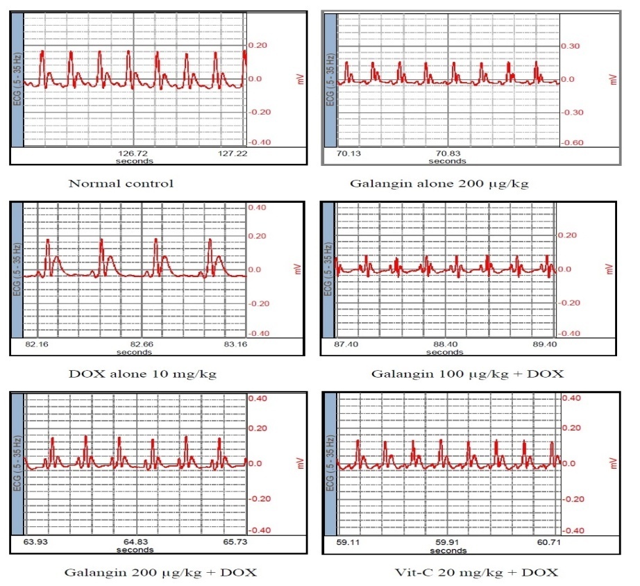
Fig. 1: Effect of Galangin on Electrocardiographic patter on DOX - induced cardio toxicity in rats
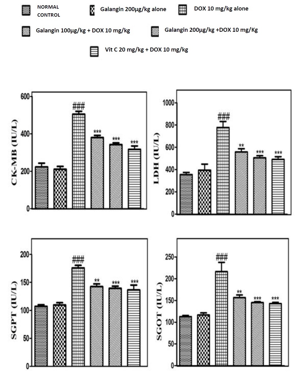
Fig. 2: Effect of Galangin in CK-MB, LDH, SGOT, SGPT on DOX - Cardio toxicity in rats. Each bar represent the Mean ± SEM (n=6), **p<0.001; ***p<0.001; compared with DOX alone rats. ***p<0.001 compared with normal control. One-way ANOVA followed by Turkeys* post test

Fig. 3: Effect of Galangin in CAT, GSH, LPO, TT on DOX - Cardio toxicity in rats. Each bar represent the Mean ± SEM (n=6), *p<0.05; **p<0.01; ***p<0.001 compared with DOX alone rats. ***p<0.001 compared with normal control. One-way ANOVA followed by Turkeys* post test
Effect of Galangin on serum enzyme markers
It is observed from figure-2, that the rats administered with DOX (10 mg/kg) shows a significantly increase in the levels of CK-MB, LDH, SGPT and SGOT as compared to Normal rats, Rats pretreated with Galangin (200µg/kg) does not showed significant change in the enzyme levels as compared with the Normal control rats, pretreated with different doses of Galangin (100 or 200µg/kg) or Vit-C(20 mg/kg) to DOX injected rats shows significant decrease (p < 0.001) in the level of CK-MB, LDH, SGPT and SGOT as compared to DOX alone treated group.
Effect of Galangin on Oxidative stress biomarkers
Reduced Glutathione, Catalase & Total Thiol
It is observed from figure-3, that the GSH, CAT and TT level in the DOX alone treated rats were significantly (p<0.001) decreased, when compared with the normal control rats. There levels in pretreated with Galangin (100 & 200µg/kg) and vit-C in DOX treated groups, were increased significantly (p<0.001) when compared to DOX alone group. Whereas GSH, CAT and TT level in Galangin alone treated rats did not modify.
Lipid Peroxidation (LPO)
It is observed from figure-3, that the TBARS level in the DOX alone treated rats were significantly (p<0.001) increased compared with the normal control rats. The TBARS level in pretreated with Galangin (100 &200µg/kg) Vit-C in DOX treated groups, were decreased significantly (p<0.01; p<0.001; p< 0.001, respectively) when compared to DOX alone group. There was no significant change in TBRS level in Galangin alone treated rats.
Histopathological changes on DOX-induced cardio toxicity
Histopathological examination of myocardial tissue as shown in figure-4, indicates that the normal control animals exhibited clear integrity of myocardial membrane. Control rats showed normal cardiac fibers without any infarction. The heart sections obtained from DOX treated animals showed abundant areas of necrosis and aggregation of acute inflammatory cells and damaged vascular spaces. Animals pretreated with Galangin 100µg/kg showed improvement in the cell integrity evidenced by decreased necrotic area and reduction in infiltration of inflammatory cells. The extent of necrosis was approximately 50% in DOX alone treated animals, which was decreased to 20% by Galangin 100µg/kg pretreatment. The Galangin 200µg/kg pretreatment showed a better protection against DOX toxicity evidenced by marked reduction of necrosis and marked decrease in infiltration of inflammatory cells and maintenance of normal integrity of the cardiac muscles. The animals treated with Vit C showed marked reversal of DOX induced Histopathological changes. However, Galangin 200µg/kg alone treated animals exhibited mild inflammation.
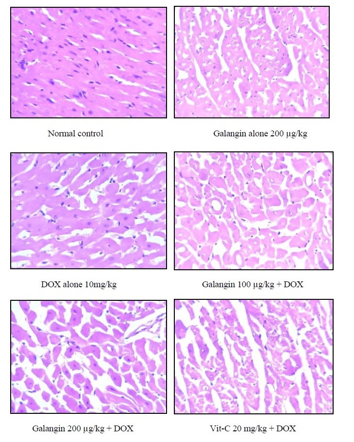
Fig. 4: Effect of Galangin: Histopathological study of heart in different groups in DOX - induced Cardio toxicity in rats
DISCUSSION
In the present study, we found that Galangin exerted a strong cardio protective effect against DOX-induced myocardial necrosis in rats. Myocardium contains an abundant amount of diagnostic marker enzymes for MI and once metabolically damaged, it releases its intracellular contents into the extracellular fluid [15]. Hence, the serum levels of these marker enzymes reflect the alterations in membrane integrity and/or permeability. Cytosolic enzymes CK-MB, LDH, SGOT and SGPT which serve as the diagnostic markers, leak out from the damaged tissue to the blood stream when cell membrane becomes permeable or ruptured [16,17]. DOX administration to rats showed elevated levels of serum CK-MB [18], which was found to be significantly low in the Galangin pretreated rats. Lactate dehydrogenase (LDH) rises within 24-48 hours after a heart attack and peaks in two to three days in the serum [19]. Consistent with the above clinical observations, in the present study we observed a significant rise in the LDH levels of rats treated with DOX after 48 or 72 h of the respective treatment. Galangin pretreatment significantly reduced the elevated levels of LDH indicating the reduction in the severity of MI.
It has been reported that infarction of as little as 10% of the total myocardium produces a significant rise in the serum levels of SGOT and is linear with the amount of infarction [20]. DOX treatment elevated these enzyme levels in serum to a significant extent. Pretreatment with Galangin (100 & 200µg/kg) or Vit-C (20mg/kg) significantly lowered the DOX-induced elevation of serum levels of these diagnostic marker enzymes. It demonstrates that Galangin could maintain membrane integrity thereby restricting the leakage of these enzymes as that of Vit-C.
Administration of supra maximal doses of DOX had been reported to induce severe oxidative stress and impair the myocardial function.[21, 22] Over production of ROS can cause severe impairment of cellular functions and necrotic lesions in the myocardium of rats. On the other hand CAT, TT, GSH constitute a mutually supportive defense team against oxidative injury.[23]
As described earlier, increase in MDA level was observed in the heart tissues after DOX administration. Malondialdehyde is a major lipid per-oxidant end product; the increased level of MDA indicates activation of the lipid per-oxidative process, resulting in irreversible damage to hearts of animals subjected to DOX stress [13,14. 23]. The results presented in this study indicated that pretreatment with Galangin (100 or 200µg/kg) could inhibit DOX induced elevation of MDA content as that of Vit-C.
The ECG is considered the single most important initial clinical test for diagnosis of myocardial ischemia and infarction. The DOX administration significantly increased ST and QT interval while the heart rate was significantly decreased as compared to control rats. The pretreatment with Galangin in DOX treated rats prevented the pathological alterations in the ECG suggestive of its cell membrane protective effect in the dose dependant manner as that as Vit-C.
The results of the serum, tissue biomarkers and ECG were well correlated with the Histopathological changes in the heart of disease and treatment groups. The myocardial tissue in control rats illustrated clear integrity of the myocardial cell membrane and absence of inflammatory cell infiltration. DOX injected rats showed coagulative necrosis, separation of cardiac muscle fibers and infiltration of inflammatory cells. The reduced inflammatory cell infiltration and normal cardiac muscle fiber architecture in Galangin treated rats further confirmed the cardio protective effect.
Augmentation of endogenous antioxidants, maintenance of the myocardial antioxidant status and significant restoration of most of the altered hemodynamic parameters may contribute to Galangin's cardio protective effect.
CONCLUSION
We conclude that the present study provides experimental evidence that Galangin has strong antioxidant activity, and it can maintain cell membrane integrity, ameliorate oxidative stress and improve cardiac systolic/diastolic dysfunction induced by high-dose DOX administration. These findings might be helpful to understand the beneficial effects of Galangin against myocardial injury although further study is needed to confirm its mechanism.
CONFLICT OF INTERESTS
Declared None
REFERENCES
- Karnik R. Soya in Heart Health. Q Med Rev 2004;55:1-35.
- Cardiovascular health; 2012. http: //www. world-heart-federation. org/cardiovascular-health/global-facts-map.
- Kumar D, Kirshenbaum LA, Li T, Danelisen I, Singal PK. Apoptosis in adriamycin cardiomyopathy and its modulation by probucol. Antioxid Redox Signaling 2001;3(1):135-45.
- Singal PK, Pierce GN. Adriamycin stimulates Ca2+ binding and lipid peroxidation but depress myocardial function. Am J Physiol 1986:250;419-24.
- Doroshow JH. Doxorubicin-induced cardiac toxicity. N Engl J Med 1991;324(12):843-5.
- Shan K, Lincoff AM, Young JB. Anthracycline-induced cardiotoxicity. Ann Intern Med 1996;125(1):47-58.
- Vijaykumar MK, Ajay GN. Antiarthritic effect of galangin isolated from rhizomes of alpinia officinarum in complete freund’s adjuvant-induced arthritis in rats. IJPPS 2014;6(4):500-5.
- Maria GM. Chemical and biological properties of propolis from the western countries of the mediterranean basin and portugal. IJPPS 2013;5(3):403-9.
- Monjur AL, Manabendra DC, Pankaj C. In silico screening of cardioprotective activity of some flavonols. IJPPS 2014;6(2):528-1.
- De Beer EL, Bottone AE, Voest EE. Doxorubicin and mechanical performance of cardiac trabeculae after acute and chronic treatment: a review. Eur J Pharmacol 2001;415(1):1-11.
- Sivakumar AS, Viswanathan P, Anuradha CV. Dose-dependent effect of galangin on fructose-mediated insulin resistance and oxidative events in rat kidney. Redox Rep 2010;15(5):224-32.
- Kaiserova H, Simunek T, van Bast A, der Vijgh WJ, Kvasnickova E. Flavonoids as protectors against doxorubicin cardiotoxicity: Role of iron chelation, antioxidant activity and inhibition of carbonyl reductase. Biochim Biophys Acta 2007;1772:1065-74.
- Bradley DW, Maynard JE, Emery G, Webster H. Transaminase activities in serum of long-term hemodialysis patients. Clin Chem 1972;18:1442.
- Wolf PL, Williams D, Coplon N, Coulson AS. Low aspartate transaminase activity in serum of patients undergoing chronic hemodialysis. Clin Chem 1972;18:567-8.
- Suchalatha S, Devi SCS. Protective effect of terminalia chebula against experimental myocardial injury induced by isoproterenol. Indian Biol 2004;42:174-8.
- Panda VS, Naik SR. Cardioprotective activity of ginkgo biloba phytosomes in isoproterenol induced myocardial necrosis in rats: a biochemical and histoarchitectural evaluation. Exp Toxicol Pathol 2008;60:397-404.
- Wakade AS, Sha AS, Kulkarni MP, Juvekar AR. Protective effect of piper longum L on oxidative stress induced injury and cellular abnormality in adriamycin induced cardiotoxicity in rats. Indian Biol 2008;46:528-33.
- Farvin KHS, Anandan R, Kumar SH, Shiny KS, Sanka TV, Thankappan TK. Effect of squalene on tissue defense system in isoproterenol-induced myocardial infarction in rats. Pharmacol Res 2004;50:231-6.
- Garba IH, Ubom GA. Total serum lactate dehydrogenase activity in acute Plasmodium falciparum malaria infection. Singapore Med J 2005;46:632-4.
- Dewar HA, Rowell NR, Smith AJ. Serum glutamic oxalacetic transaminase in acute myocardial infarction. Brt Med J 1958;8:1121-5.
- Wang SB, Tian S, Yang F, Yang HG, Yang XY, Du G, et al. Hua Cardioprotective effect of Salvianolic acid A on isoproterenol-induced myocardial infarction in rats. Eur 2009;615:125-32.
- Ayaz SA, Bhandari U, Pillai KK, Indian J. Influence of DL α-lipoic acid and vitamin-E against doxorubicin-induced biochemical and histological changes in the cardiac tissue of rats. Pharmacol 2005;37:294-99.
- Kumar P, Muralinath E, Swamy P, Krishna V, Rutin M. Raja Lakshmana hari potential action of vitamin-e, quercetin against the doxorubicin-induced cardiomyopathy. IJPPS 2011;3(4):280-4.